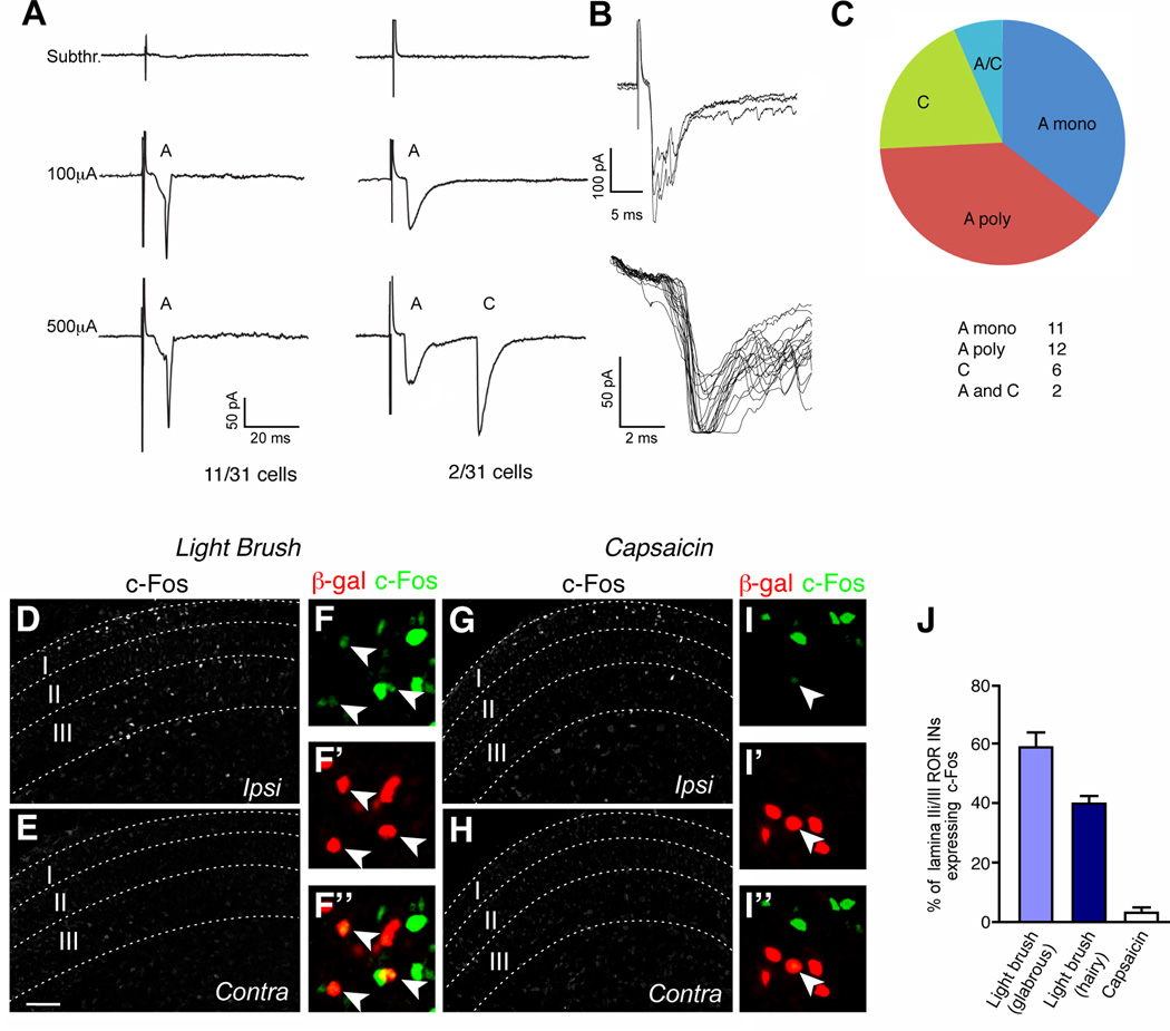
Figure 4. RORα INs are functionally innervated by low threshold mechanosensory afferents.
(A) Example of a RORα IN in L4 displaying an A fiber monosynaptic current triggered by low intensity stimulus. Monosynaptic connectivity was confirmed by a 2Hz frequency stimulation. (B) Example of a RORα IN with dual A and C inputs. (C) Classification of potentials in RORα INs induced by dorsal root stimulation. n = 31 cells. (D–I) Sections through the lumbar spinal cord of P6 RORαCre; R26ds-HTB mice stained with antibodies to c-Fos and β-gal after light brushing (D–F) or injecting capsaicin into the footpad (G–I). Arrowheads in F and I indicate c-Fos+ RORα INs. Scale bars: 50 µm. Panels E and H show the contralateral side. (J) Fraction of RORα INs (β-gal+) coexpressing c-Fos. Only lamina IIi/III cells located within the domain displaying elevated c-Fos expression were counted. Cell counts of RORα INs exhibiting c-Fos immunoreactivity: brush stimulation of glabrous skin, 1593/2830 RORα cells; brush stimulation of hairy skin, 834/2103 RORα cells; capsaicin injection, 20/711 RORα cells. n = 4 animals for each experiment. Data: mean ± SEM. Scale bar: 100 µm (D,E,G,H).

