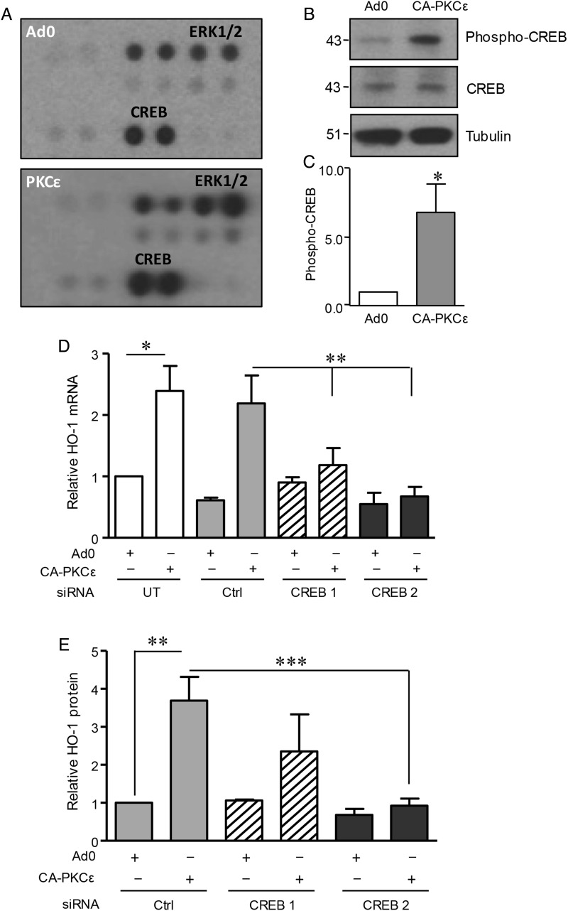
Figure 3.
Role of CREB1 in PKCε-mediated HO-1 induction. (A) HUVECs were transfected with CA-PKCε or Ad0 (MOI 100) for 16 h prior to generation of lysates to probe a phosphokinase Ab array. Results for the phosphorylation of ERK1/2 and CREB1 are shown and are representative of two separate experiments. (B) HUVECs were transfected with CA-PKCε or Ad0 (MOI 100) for 16 h and immunoblotted for phospho-CREB(Ser133), CREB1, and tubulin, with (C) a histogram showing pooled quantification data from five experiments. (D) HUVECs were left UT or transfected with a single CREB1 siRNA oligo (CREB1), pooled CREB1 siRNA oligos (CREB2), or control (Ctrl) siRNA (40 nM) for 24 h prior to transfection with control Ad0 or CA-PKCε adenovirus (MOI 100) for 16 h. HUVECs were lysed and HO-1 mRNA levels analysed by qRT-PCR. (E) HUVECs were transfected as above with Ctrl, CREB1, or pooled CREB1 siRNAs, prior to analysis of HO-1 by immunoblotting. The figure shows a representative blot. Pooled HO-1 quantification data from at least four individual experiments were corrected for tubulin expression where appropriate. Data are presented normalized to control-transfected cells (mean ± SEM). One-sample t-test (C), ANOVA (D and E), *P < 0.05, **P < 0.01, ***P < 0.001.

