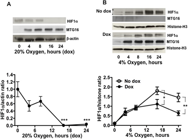
Fig 7. MTG16 reduces the level of HIF1a.
A. The level of HIF1α was examined by Western blotting in lysates from Raji-MTG16 cells incubated under exposure to 20% O2 with 20 ng/ml of doxycycline to induce synthesis of MTG16 (as detected by α-MTG). HIF1α becomes undetectable after 8 to 16 h of incubation. Blotting data from one out of three experiments is shown (top). The intensity of the bands was quantified by densitometry and expressed as HIF1α/β-actin ratio, n = 3. The level of HIF1α was reduced after 16 h; n = 3, ***P<0.001(bottom). B. The level of HIF1α was examined by Western blotting using the α-MTG antibody in lysates from Raji-MTG16 cells incubated under hypoxia with 4% oxygen with or without 20 ng/ml of doxycycline to induce synthesis of MTG16. Blotting data from one out of three experiments is shown (top). To compensate for a lower level of HIF1α at normoxia compared to hypoxia, the exposure time of photographic film was 60 sec for experiments performed at 20% O2 (A) compared to 20 sec for experiments carried out at 4% O2 (B). The intensity of the bands was quantified by densitometry and expressed as HIF1α/β-actin ratio. The level of HIF1α was reduced after 24 h; n = 3, **P<0.01(bottom).

