
Fig 6. Focal activity within insula cortex correlating with physiological changes.
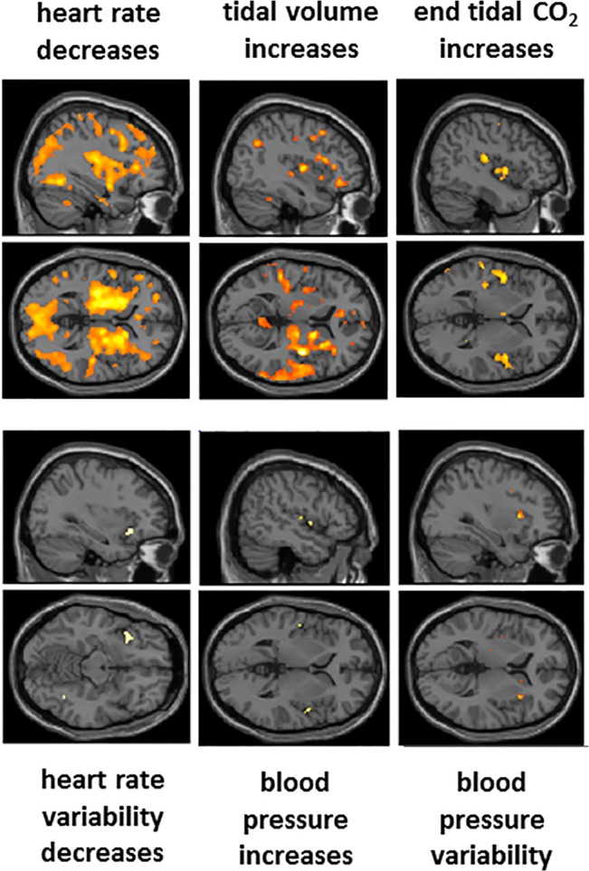
Sagittal and horizontal sections from a standard template brain presented to highlight the location of activity changes within insula cortex associated with task-induced changes in physiological regressors. Of note are the deep insula components of signal change associated with heart rate decrease, merging with a marked engagement of basal ganglia, mirroring striatocortical activation previously observed in relation to expectancy-related heart rate deceleration [22]. The broadly distributed signal change, associated with this particular analysis arguably appears to carry most artefact, beyond what was controlled for by including movement, global and arterial O2 / end tidalCo2 regressors as confounding covariates within the analyses. Ventilation (VT) evoked predominantly right hemispheric changes in parietal and insula cortices and basal ganglia. Increases in end tidal CO2 was associated with enhanced activation within posterior ‘primary interoceptive’ insula, but did not impact global signal at threshold significance. Heart rate variability, blood pressure increases and blood pressure variability were associated with focal activation of distinct subregions of anterior and mid insula consistent with viscerotopography [23]. Data is illustrated at a significance of P<0.05 corrected, determined from the combination of voxel-wise significance and a cluster extent thresholds (see Methods).