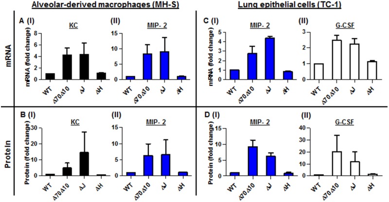
Fig 4. In vitro infection of alveolar macrophages and lung epithelial cells with Y. pestis strains.
In vitro infection of MH-S alveolar-derived macrophages (A-B), and TC-1 lung-derived epithelial cell lines (C-D) with 50 MOI of the virulent Y. pestis strain Kim53 (WT) and its Yop-depleted derivatives Kim53ΔYopJ (ΔJ), Kim53ΔYopH (ΔH) and Kim53Δ70Δ10 (Δ70Δ10). The mRNA (A and C) and protein (B and D) levels were quantified using qPCR and ELISA, and are presented as the fold change relative to the WT-Kim53 Y. pestis strain.

