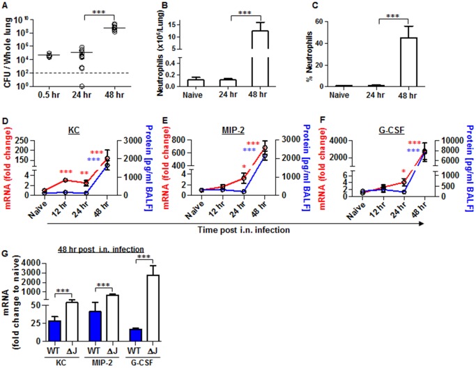
Fig 5. Neutrophils homing to lungs and bacterial propagation following airway infection of mice with the YopJ-deleted strain (Kim53ΔYopJ).
Bacterial propagation in the lungs of C57BL/6J mice infected i.n. with 1x10⁵ cfu of the mutated Y. pestis strain Kim53ΔYopJ was determined by plating the samples and calculating the cfu. Absolute numbers (B) and percentages (C) of neutrophils in the lungs of the infected mice at the indicated time points post infection were determined by flow cytometry analysis as described in Fig 1. Whole lung extracts or BALF were collected at the indicated time points post i.n. infection with 1x10⁵ cfu of Y. pestis strain Kim53ΔYopJ and then analyzed for the mRNA expression (red line) and protein level (blue line) of KC (D), MIP-2 (E) and G-CSF (F). (G) Comparison between the mRNA levels of KC, MIP-2 and G-CSF in the lungs of C57BL/6J mice at 48 hr post i.n. infection with 1x10⁵ cfu of Kim53 (WT) or Kim53ΔYopJ (ΔJ). Fold changes were determined in comparison to naïve mice. The results are presented as the means ± SEM (*p<0.05, **p<0.01, ***p<0.001). n = 6–12 mice per group of at least 3 independent experiments.

