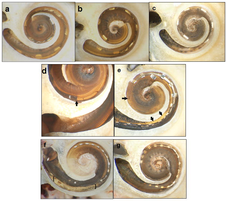
Figure 4.

Histologic analysis with bone of promontory removed and basilar membrane (BM) in place. (a–c) Specimens 1–3 (Medel standard array electrodes) showed no intracochlear trauma, but incomplete insertion with 10, 8, and 8 electrodes intracochlear. (d–e) Specimens 4 and 5 (Advanced Bionics HiFocus 1J) had contrasting results with minimal trauma noted in Specimen 4 (d) where the tip of the electrode (marked as E) is noted with the dark and a subtle tear of BM near the cochleostomy site was identifiable (difficult to appreciate in 2-D photographs). Specimen 5 (e) had significant trauma with penetration of the osseous spiral lamina (OSL) and insertion of the electrode completely in scala vestibuli; the arrows to the right indicate the OSL injury and the horizontal arrow indicates the tip in scala vestibule resting on the BM. (f–g) Specimens 6 and 7 were Cochlear SRA electrodes. Specimen 6 (f) had a significant fracture in the OSL just distal to the cochleostomy (starting point noted by the diagonal arrow) and subsequent injury to the BM in the area between the brackets. Specimen 7 (g) had a small tear in the BM just anterior to the round window.
