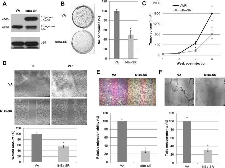
Fig 2. Inactivation of p65 reduces the abilities of in vitro colony formation, in vivo tumor formation, migration, invasion, and angiogenesis in NPC.
(A) Western blot analysis shows the expression of exogenous IκBα-SR with triple HA-tag (^) in HONE1 cells. The p84 served as a loading control. (B) 2D colony formation assay shows that IκBα-SR suppressed the colony-forming ability of HONE1 cells compared to pWPI-vector alone (VA). Bar graphs indicate data obtained from an average of triplicate experiments ± S.E.M. (C) Nude mice were inoculated subcutaneously with IκBα-SR and pWPI-VA HONE1 cells. IκBα-SR-transduced cells showed delayed and reduced tumor growth kinetics compared to pWPI-VA. Each data point represents an average tumor volume of six injection sites inoculated for each cell population ± S.E.M. (D) Wound healing analysis for pWPI-VA and IκBα-SR showed delayed migration of IκBα-SR cells compared to VA. Bar graphs show the percentage difference between IκBα-SR and VA cells ± S.E.M. (E) Migration chamber assays showed that IκBα-SR-expressing HONE1 cells reduced migration compared to VA cells. Data represented on the bar graph are the average of triplicate experiments ± S.E.M. (F) HUVEC tube formation was suppressed with IκBα-SR conditioned medium compared to that of VA cells. Data represented on the bar graph are the average of triplicate experiments ± S.E.M. The (*) for all graphs indicate P-value < 0.05.

