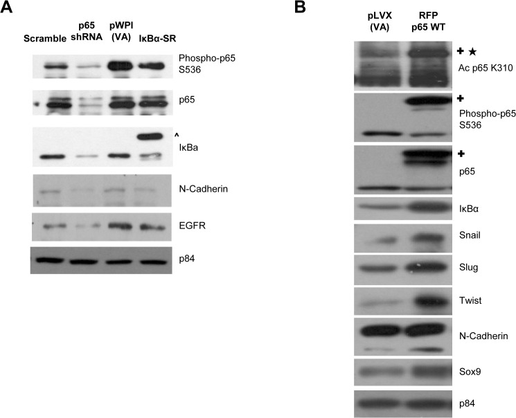
Fig 4. p65 signaling pathway regulates the protein expression of EGFR and EMT markers.
(A) Western blot analysis of p65shRNA- HONE1 cells showed reduced levels of phosphorylated p65 S536, total p65, and total IκBα compared to scramble control. IκBα-SR- HONE1 cells only showed a decreased amount of phosphorylated p65 S536, but did not affect the overall amount of total p65 when compared to pWPI-VA. Endogenous and exogenous IκBα are labeled as shown. The p84 was used as a loading control for scramble and p65 shRNA, pWPI-VA and IκBα-SR, separately. Protein expression of N-cadherin and total EGFR were all reduced in p65 shRNA and IκBα-SR HONE1 cells compared to scramble and pWPI-VA control. The (^) indicates the exogenous 3xHA-IκBα-SR. (B) Western blot analysis of p65 WT-overexpressing HONE1 cells showed increased acetylation at K310 and enhanced phosphorylation at S536 compared to VA cells. The p65 WT overexpression increases the protein levels of IκBα, snail, slug, twist, N-cadherin, and sox9, compared to VA cells. The p84 was used as a loading control. The (✚) indicates the exogenous RFP-p65 WT. The (★) indicates the acetylation band in the exogenously expressed RFP-p65 WT.

