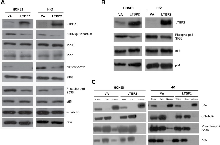
Fig 5. LTBP2 regulates the p65 signaling pathway.
(A) Phosphorylated IKKα/β S176/180 and phosphorylated IκBα SS32/36 are also reduced in LTBP2-infected cells. Phosphorylated p65 serine 536 level is reduced in LTBP2-infected cells compared to VA in HONE1 and HK1. The p84 and α-tubulin were used as loading controls. (B) Matrigel plug tumors show a reduction in phosphorylated p65 S536 in LTBP2-transduced cells compared to VA. The p84 was used as loading control. (C) Subcellular fractionation results show that phosphorylated p65 S536 was reduced in the nuclear fraction of LTBP2-transduced cells compared to VA. The p84 was used as control to determine nuclear fraction, while α-tubulin was used as the control for the cytoplasmic fraction.

