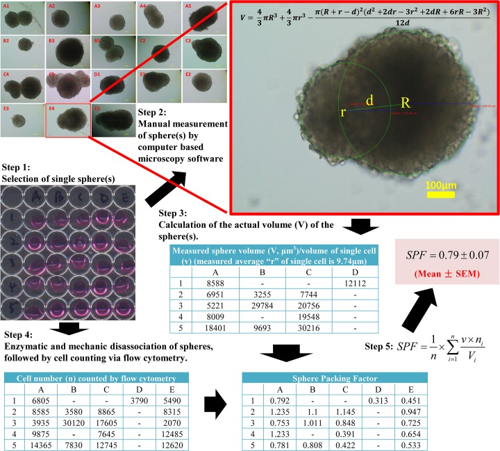
Fig 3. A general protocol for SPF calculation using H1299 TS.
Step 1: Spheres were harvested and seeded individually in each well. Step 2: The diameter of spheres was measured by computer-based imaging software, and documented. Note that the three labels in red are software generated measurement of distance between centers of two spheres (223.08 μm), radius of left sphere (165.45 μm) and radius of right sphere (248.03 μm), respectively (from left to right). Step 3: The volume of each sphere was calculated based on measured diameter obtained from step 2. Step 4: Spheres were disassociated enzymatically and mechanically to make single cell suspension, followed by precise cell counting by a cytometry. Meanwhile, the diameter of single floating cells from spheres was measured. Step 5: SPF was calculated by an indicated formula.

