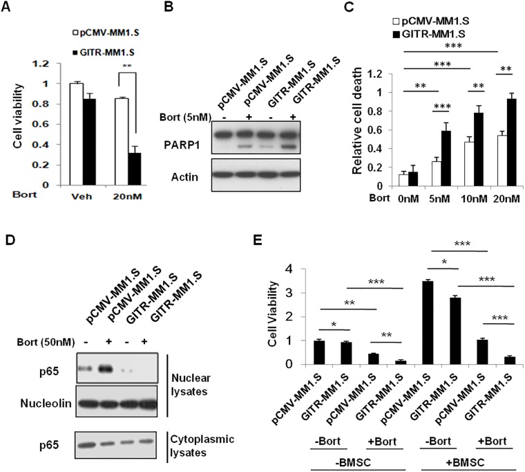
Fig 5. Overexpression of GITR enhanced sensitivity to Bortezomib-induced apoptosis in MM1.S cells.
A. Empty vector and GITR-transfected MM1.S cells were treated with Bortezomib for 48 hours. Cell viability was assessed by CellTiter-Glo assay. Data represent mean ± SD, **P<0.01 compared with indicated groups. B. Empty control and GITR expressing MM1.S cells were exposed to different doses of Bortezomib and incubated overnight. Cells were lysed and subjected to Immunoblotting using anti-PARP1 and Actin antibodies. C. Empty control and GITR expressing MM1.S cells were exposed to different doses of Bortezomib and incubated for 24 hours. The number of dead cells were assessed by PI single staining and quantified by Flowjo software. Data represent mean ± SD, **P<0.01 and ***P<0.001 compared with indicated groups. D. NF-κB activity was evaluated in control and GITR expressing MM1.S cells. Nuclear protein lysates were subjected to western blot using anti-p65 and nucleolin antibodies. E. Empty vector and GITR-transfected MM1.S cells were treated with 50nM Bortezomib for 48 hours in co-cultured with or without BMSC. After 48 hours incubation, the cell viability was assessed by CellTiter-Glo assay. Data represent mean ± SD, *P<0.05, **P<0.01 and ***P<0.001 compared with indicated groups.

