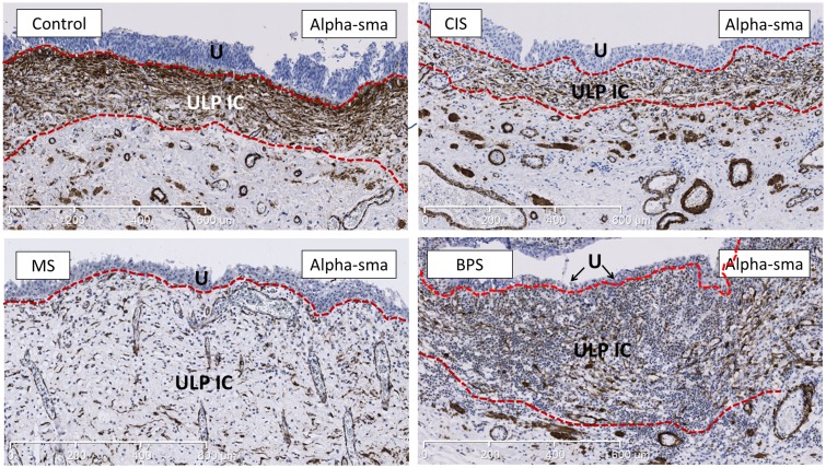
Fig 2. Representative images of staining for alpha-sma (marker for ULP IC) showing the increased thickness of the alpha-sma+ ULP IC area in MS and BPS bladders.
The ULP IC area lies between the 2 dotted red lines. The alpha-sma+ cell-types under the ULP IC area are MM-fibres and perivascular smooth muscle cells (which were excluded from analysis); IC in the DLP are negative for alpha-sma. In BPS bladder heavy inflammatory infiltrate is laying in between the IC, while in MS bladder this infiltrate is less pronounced. U is urothelium, ULP IC equals upper lamina propria interstitial cell. Scale bars indicate 600μm.

