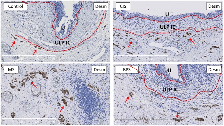
Fig 4. Representative images of desmin-staining (marker for MM) showing the increased development of desmin+ MM in MS bladders compared to controls.
MM is more developed in MS bladders, both vertically and horizontally. MM (indicated by red arrows) lies just below the ULP IC area (between the 2 dotted red lines). In BPS and CIS bladders there was a trend towards increased MM development, but no significant difference was found (see Fig 3). Scale bars indicate 600μm. U is urothelium, ULP IC equals upper lamina propria interstitial cells.

