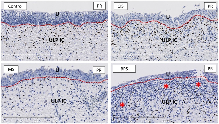
Fig 7. Representative images of PR-staining, showing the decreased amount of PR+ ULP IC in MS bladders.
PR shows a nuclear expression and is widely expressed in ULP IC. Asterisks indicate heavy inflammatory infiltrate in the ULP area in BPS. U is urothelium, ULP IC equals upper lamina propria interstitial cells (red dotted line separates urothelium form the ULP IC area). Scale bars indicate 300μm.

