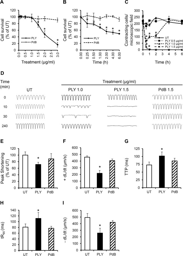
Fig 4. PLY at sub-lytic doses adversely affect cardiomyocyte function in vitro.
(A) Viability of HL-1 cells was assessed 30 min after incubation with increasing concentrations of PLY and PdB using the WST-8 assay. Viability of untreated cells (UT) was set at 100%. Data are presented as Mean±SD. * p<0.05 (n = 4). (B) Time course of HL-1 cell viability after incubation with 1.5 μg/ml PLY or PdB. *p<0.05 (n = 3). (C) Effects of increasing concentrations of PLY on the total number of spontaneously contracting HL-1 cells over time. Data are presented as Mean±SD. *p<0.05 (n = 4). (D) Representative traces of cardiomyocyte contraction before and after PLY and PdB treatment (n = 4). (E-I) Effects of PLY and PdB (1.0 μg/ml) on Peak Shortening (E), +dL/dt (F), TTP (G), tR90 (H) and -dL/dt (I) of HL-1 cells after 30 min treatment are presented as Mean±SD. *p<0.05 (n = 9 from 3 independent experiments).

