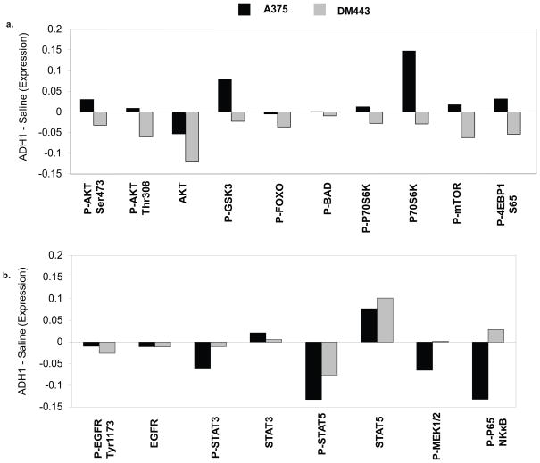
Figure 3. Comparative effects of ADH-1 treatment on signaling pathways in the A375 and DM443.
RPPA analysis was performed on xenografts of the A375 (Black bars) and the DM443 (Gray bars) harvested one hour after treatment with saline or ADH-1 as outlined in Figure 1. The effects of ADH-1 treatment were determined by subtracting the exponential value of the relative expression for each of the indicated proteins following saline treatment from the observed expression following ADH-1 treatment. Proteins with a relative change of >0 were increased with ADH-1 treatment; <0 indicates inhibition with ADH-1 treatment. (a.), expression of total- and phospho-proteins in the PI3K-AKT pathway. (b.) expression of total and phospho-proteins in other pro-survival kinase signaling pathways.

