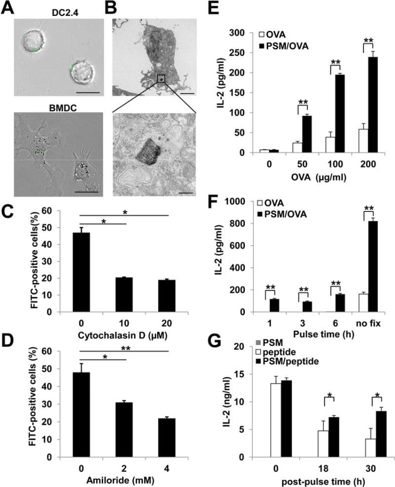
Figure 1.

PSM-loaded antigen was efficiently internalized by dendritic cells through phagocytosis and macropinocytosis.
(A) Representative microscopic views of DCs internalizing PSM particles loaded with OVA-FITC nanoliposomes. Upper panel, DC2.4 cells; lower panel, bone marrow-derived DCs. Scale bar, 25 μm.
(B) A representative transmission electron microscopy picture showing the vesicular structure around a PSM/OVA particle inside the DC. Upper panel, 5,000 X; lower panel, 50,000 X. Upper panel scale bar, 4 μm; lower panel bar, 0.5 μm.
(C) DC uptake of PSM/FITC-OVA in the presence of phagocytosis inhibitor cytochalasin D.
(D) DC uptake of PSM/FITC-OVA in the presence of macropinocytosis inhibitor amiloride.
(E) IL-2 production by B3Z cells after co-incubation with DCs pre-treated for 3 h with different concentrations of soluble OVA or PSM/OVA.
(F) IL-2 production by B3Z cells after co-incubation with DCs that were primed with 50 μg/ml soluble OVA or PSM/OVA followed by fixation at different time points.
(G) IL-2 production by B3Z cells after co-incubation with DCs. The DCs were extensively washed after priming with 5 μg/ml soluble peptide or PSM/peptides, and either immediately co-incubated with B3Z cells (0 h), or cultured for 18 to 30 h before T cell co-incubation.
Data are presented as mean ± S.D. *, P<0.05, **, P<0.01. See also Figure S1.
