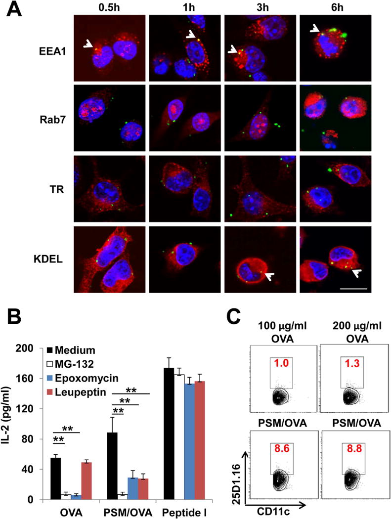
Figure 2.

Antigen presentation of PSM-loaded OVA.
(A) Subcellular transport of antigen. DCs were incubated with PSM/FITC-OVA for 0.5 h, and then washed extensively to remove unbound particles. The cells were cultured for an additional 0.5 h, 1 h, 3 h, or 6 h before fixation followed by immunostaining with antibodies recognizing specific organelles (EEA1 for early endosome, Rab7 for late endosome, TR for recycling endosome, and KDEL for the ER). Scale bar, 25 μm.
(B) IL-2 production by B3Z cells after co-incubation with DCs treated with 100 μg/ml OVA or PSM/OVA in the presence of proteosome inhibitors (MG-132, epxomycin) or lysosome inhibitor (leupeptin). Peptide I (100 ng/ml) served as the positive control.
(C) OVA cross-presentation by BMDCs. BMDCs were incubated with OVA or PSM/OVA for 16 h, followed by harvest and labeling with the anti-CD11c antibody identifying DCs and the 25D1.16 antibody recognizing OVA257–264/H-2Kb complex on DC surface. Percentages of 25D1.16 staining positive populations in DCs were shown in red numbers.
Data are presented as mean ± S.D. *, P<0.05, **, P<0.01. See also Figure S2.
