
Figure 3.
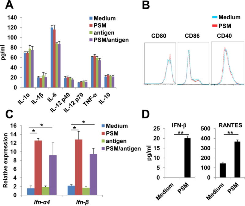
PSM induced IFN-I signaling in DCs.
(A) Protein levels of pro-inflammatory cytokines in culture media of BMDCs 24 h after incubation with PSM, free antigen, or PSM/antigen. Cell culture medium served as the negative control.
(B) Expression pattern of co-stimulatory molecules on the surface of BMDCs 24 h after incubation with PSM.
(C) QPCR analysis on mRNA levels of the Ifn-α4 and Ifn-β genes in BMDCs 5 h after co-incubation with PSM, free antigen, or PSM/OVA antigen.
(D) ELISA assay for IFN-β and RANTES in culture media of BMDCs 24 h after incubation with PSM.
Data are presented as mean ± S.D. *, P<0.05, **, P<0.01. See also Figure S3.