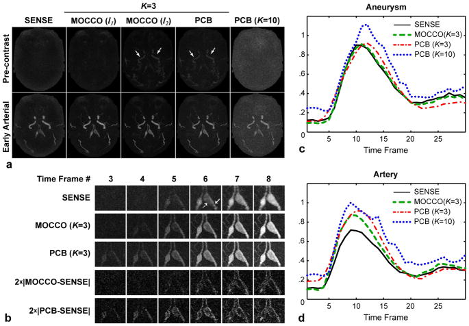
Figure 7.
Reconstruction results for intracranial CE MRA exam. a: A pre-contrast (top) and early arterial (bottom) time frames. b: Magnified ROI in the time series reconstructed by SENSE, MOCCO, and PCB (K=3), and corresponding image differences. c,d: Contrast enhancement waveforms measured in the aneurysm and its feeding artery indicated by dashed and solid arrows in (b), respectively.

