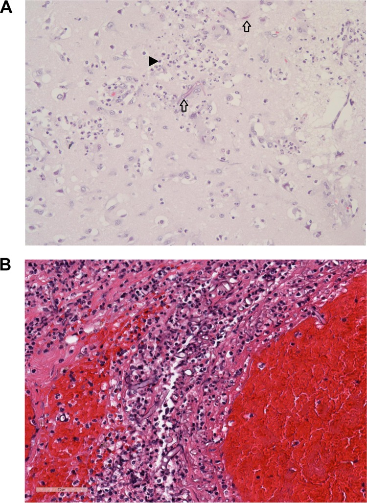
FIG 2.
(A) Tissue taken from the cortex of the right occipital lobe reveals brain parenchyma with patchy infiltration by broad, aseptate fungal hyphae (arrows) and robust acute encephalitis, as noted by the large number of neutrophils (arrowhead). No other foci of necrosis or inflammation were noted in the brain. (B) Hematoxylin-and-eosin-stained right testis and soft tissue (PDOI 42/autopsy) with acute and chronic inflammation with hemorrhage. Broad, aseptate hyphae are seen interspersed with the inflammatory cells.

