FIG 1.

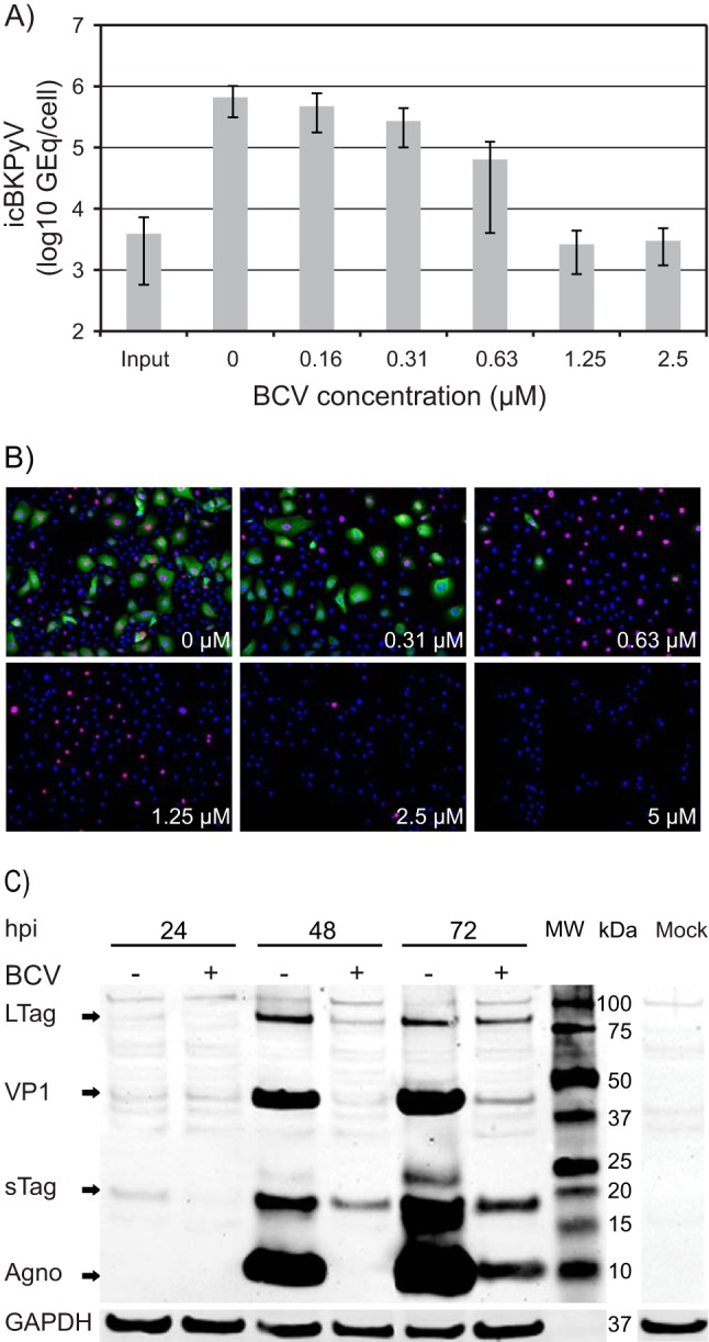
Effect of brincidofovir (BCV) on BKPyV replication in HUCs. (A) Effect of BCV on BKPyV DNA replication. Cells were seeded at 20,000 cells/cm2 and infected 24 h later with purified BKPyV at 2 FFU/cell. Infected cells, treated with the indicated concentrations of BCV, were harvested 72 hpi (70 hpt), and icBKPyV (number of genome equivalents [Geq]/cell) was measured by qPCR. The mean icBKPyV values from three independent experiments conducted and analyzed in triplicate (27 quantification reactions per mean) are presented. Error bars indicate the standard deviations of means between experiments. (B) Effect of BCV on BKPyV expression of early and late proteins at 72 hpi. Immunofluorescence micrographs of BKPyV-infected HUCs treated with the indicated BCV concentrations are shown. The cells were fixed and subsequently stained using polyclonal rabbit antiagnoprotein serum (green) for visualization of agnoprotein and the SV40 LTag monoclonal Pab416 for the visualization of early LTag (red). Cell nuclei (blue) were stained with DRAQ5. Pictures were taken with a fluorescence microscope (10× objective). (C) Effect of BCV on temporal expression of BKPyV early and late proteins. Western blot of lysates of BKPyV infected HUCs treated with 0.63 μM BCV (+) and buffer only (−). Treatment was initiated 2 h after infection, and cells were lysed at the indicated times postinfection. Electrophoresis was performed with 5.2 μg total protein per well. The membrane was stained with polyclonal rabbit anti-LTag serum, anti-agnoprotein, and anti-VP1 serum and with a mouse monoclonal antibody directed against the housekeeping protein glyceraldehyde-3-phosphate dehydrogenase (GAPDH). Lane MW, molecular weight markers.
