
FIG 4.
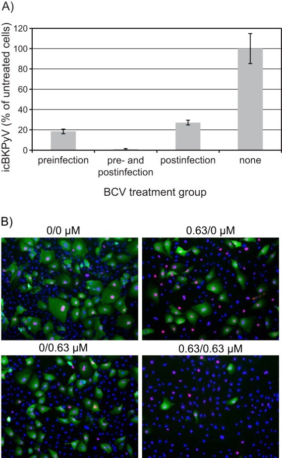
Effect of pre- and postinfection treatment with BCV. HUCs were seeded at 20,000 cells/cm2, incubated for 24 h, and then treated with 0.63 μM BCV or buffer only for 24 h prior to infection with purified BKPyV at 2 FFU/cell for 2 h. Following infection, the cells were washed with medium and then replenished with medium containing 0.63 μM BCV or buffer only. Cells were harvested for extraction of DNA (A) or fixed at 72 hpi (B). (A) The icBKPyV is plotted as a percentage of that in wells treated with buffer only (100%). Bars represent means from 3 individual experiments conducted and quantified in triplicate, giving 27 quantification reactions per mean. Error bars represent standard deviations. (B) Immunofluorescence micrographs of HUCs fixed at 72 hpi and stained using polyclonal rabbit anti-agnoprotein serum (green) for visualization of agnoprotein and the SV40 LTag monoclonal Pab416 for the visualization of early LTag (red). Cell nuclei (blue) were stained with DRAQ5. Pictures were taken with a fluorescence microscope (10× objective).