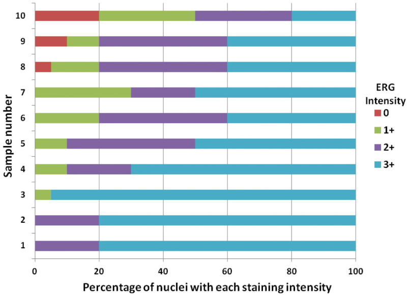
Figure 2. Analysis of ERG expression in prostate cancers by visual quantitation.
Ten tumors from radical prostatectomy specimens with ERG staining by IHC were scored for staining intensity in cancer nuclei (0, 1+, 2+ or 3+). The percentage of nuclei with each staining intensity was estimated to the nearest 5%.

