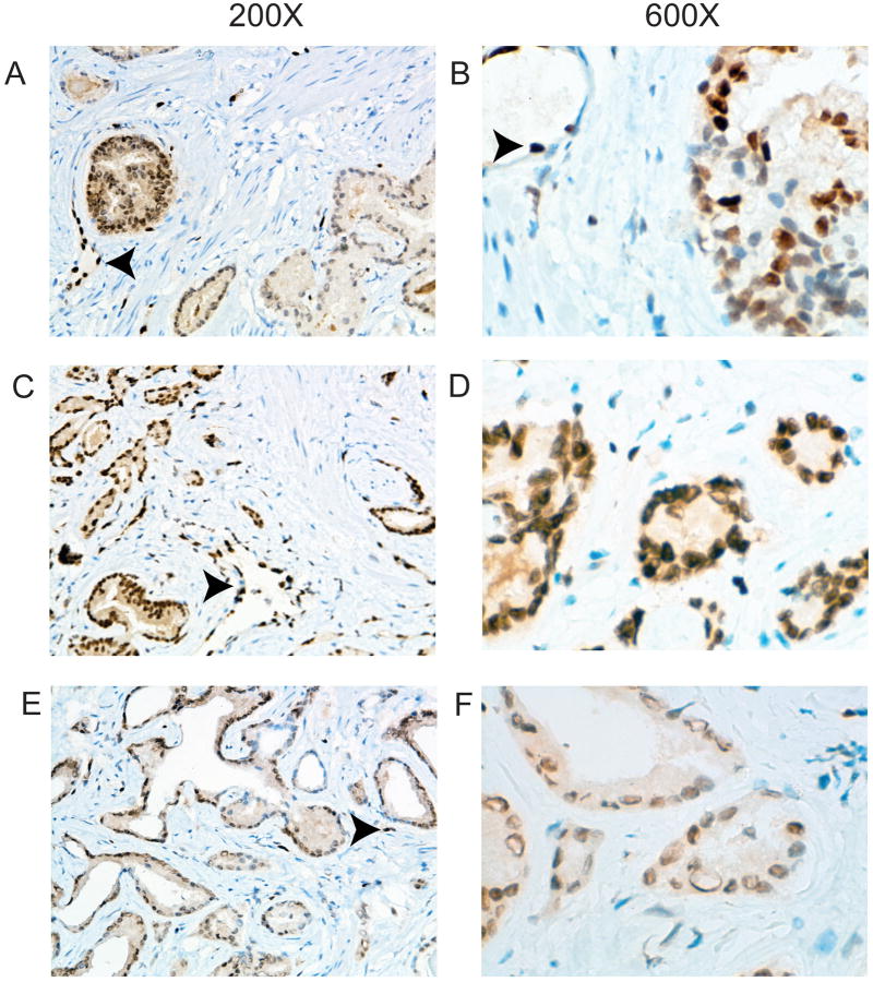
Figure 3. Immunohistochemistry with anti-ERG antibody using rapidly fixed prostatectomy tissues.
Prostate cancer tissues were snap frozen in liquid nitrogen within 15 minutes of surgery and a thin slice taken that was then fixed overnight in formalin, embedded and ERG IHC performed. A. Intrafocal variability. Weakly staining tumor is on the right with strongly staining tumor on the left. A blood vessel is indicated by the arrowhead. B. Intrafocal variability. A tumor with variable staining within a single focus ranging from negative to strong. An endothelial cell with strong staining is indicated by the arrowhead. C-F. Interfocal variability. C and D show medium and high power views of a cancer with uniform strong staining similar in intensity to endothelial cells. E and F show a cancer with medium staining. Endothelial cells are indicated by the arrowheads. Magnification is shown at top.

