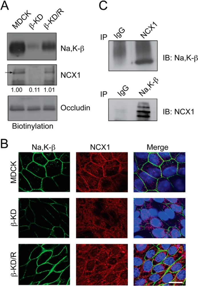
FIGURE 2.

Na,K-β associates with NCX1 and its knockdown reduces NCX1 cell surface localization. A, cell surface proteins were pulled down with streptavidin beads and immunoblotted for indicated proteins. Immunoblots show cell surface levels of Na,K-β, NCX1, and occludin in MDCK, β-KD, and β-KD/R cells. NCX1 cell surface levels expressed, as -fold change with respect to MDCK cells, are shown below the blot. The reduction in NCX1 cell surface levels in β-KD cells is statistically significant (p = 0.003). B, representative images showing immunofluorescence staining of Na,K-β (green) and NCX1 (red) in MDCK, β-KD, and β-KD/R cells. The TOPRO-3 stained nuclei are shown in blue. Scale bar = 25 mm. C, immunoblots demonstrating association of NCX1 and Na,K-β by co-immunoprecipitation (IP) analysis in MDCK cells. Representative blots from three independent experiments are shown.
