FIGURE 4.

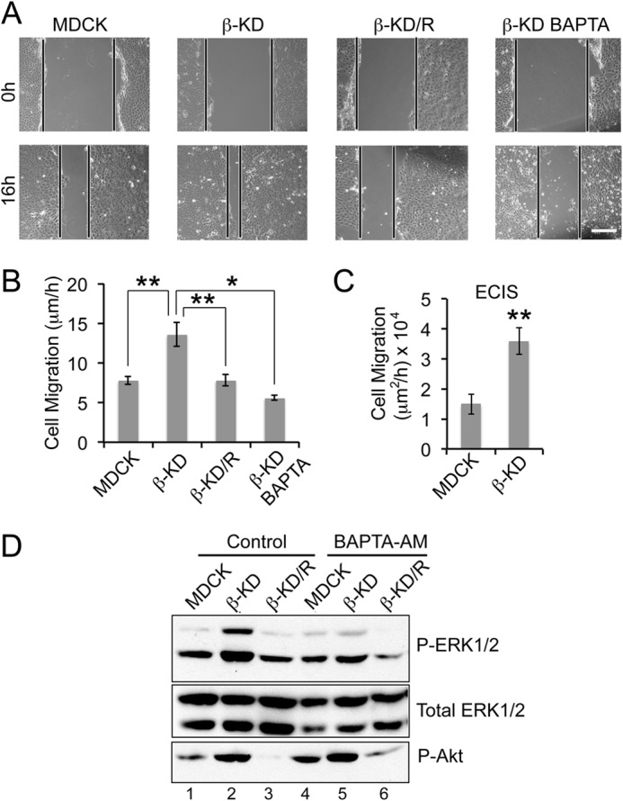
The role of Ca2+ in inducing cell migration and ERK1/2 phosphorylation in β-KD cells. A, representative images of the wound at 0 h and after 16 h in wound healing assay. Similar images were used to calculate the distance of migration over 16 h. Scale bar = 100 μm. B, the graph represents the average rate of migration calculated from three independent experiments in triplicate. Error bars denote S.E., and the asterisks indicate statistical significance (*, p < 0.05; **, p < 0.005). C, the graph represents the rate of migration obtained by ECIS wound healing assay from three independent experiments. Error bars denote S.E., and the asterisks indicate statistical significance (**, p < 0.005). D, immunoblots show the phosphorylation status of ERK1/2 and Akt (Ser-473) and total ERK1/2 (loading control) in the indicated cell lines under control conditions or with 10 μm BAPTA-AM treatment. Representative blots from four independent experiments are shown.
