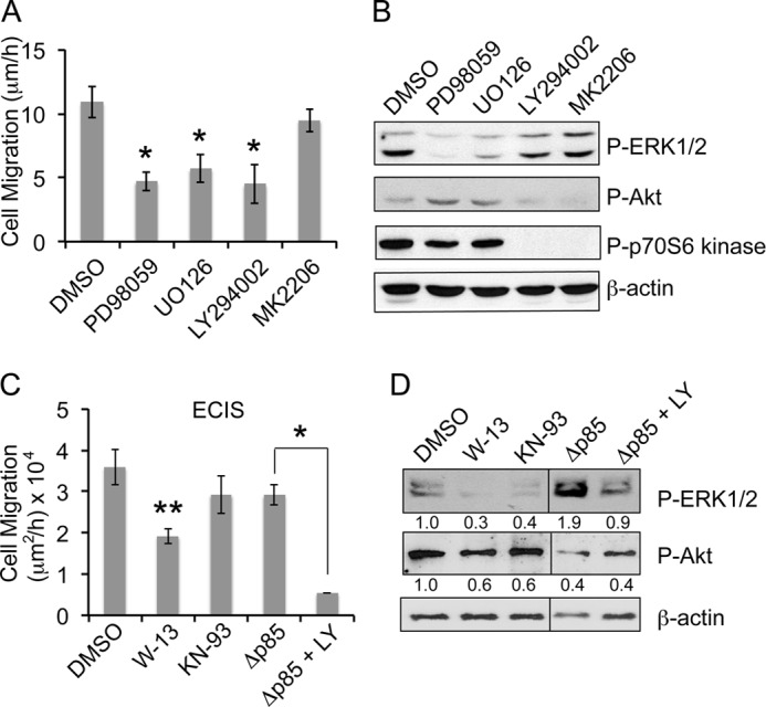
FIGURE 5.

Increased migration in β-KD cells is dependent on calmodulin/PI3K-mediated ERK1/2 activation. A, the graph shows the average rate of migration of β-KD cells treated with the indicated inhibitors (LY294002, U0126, PD98059, MK2206 at 10 μm each) from three independent experiments performed in triplicate. Error bars denote S.E. Treatment with all inhibitors except MK2206 attained statistical significance (*, p < 0.05). B, the immunoblots show the levels of phospho-ERK1/2, phospho-Akt, phospho p70S6 kinase and β-actin (loading control) after inhibitor treatment for 16 h in β-KD cells. Representative blots from three independent experiments are shown. C, the graph shows the average rate of migration by ECIS wound healing assay in β-KD cells treated with DMSO, 30 μm W-13 or 30 μm KN-93, and β-KD/ΔP85 cells after treatment with DMSO or 10 μm LY294002 from three independent experiments. Error bars denote S.E. Asterisk(s) indicate statistical significance (*, p < 0.05; **, p < 0.005). D, the immunoblots show the levels of phospho-ERK1/2, phospho-Akt, and β-actin after inhibitor treatment for 16 h. Quantification from three independent experiments expressed as -fold change normalized to β-actin loading control are indicated below the blot.
