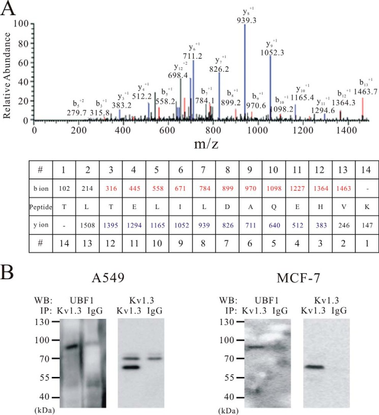
FIGURE 5.
Co-assembly of UBF1 and nuclear Kv1.3 channel. A, MS/MS spectra of the UBF1-specific peptide obtained by mass spectrometry. The b ions originate from the cleavage of the peptide backbone with N-terminal charge retention, and the y ions indicate peptide fragments with C-terminal charge retention. The b and y ions matched to the UBF1 peptide are indicated in red and blue, respectively. B, co-immunoprecipitation of Kv1.3 with UBF1 from A549 (n = 3) and MCF-7 (n = 3) nuclear extracts. IgG was used as a negative control. WB, Western blot; IP, immunoprecipitation.

