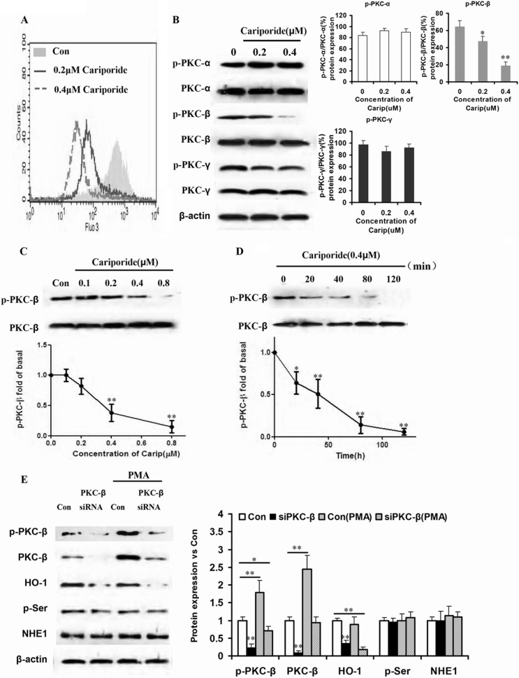
FIGURE 3.
PKC-β phosphorylation mediated HO-1 expression induced by NHE1 hyperfunction. A, K562R cells were treated by different doses of cariporide (0, 0.2, and 0.4 μm) for 20 min, and intracellular Ca2+ was detected by flow cytometry and shown in the overlap style. Con, control. B, the phosphorylation levels of classical PKC family proteins were assessed by Western blot after K562R cells were treated by cariporide (Carip). The K562R cell line was incubated with cariporide at the same concentrations, and HO-1 expression was detected by Western blot. The box and whisker plots were made to assess mRNA and protein expressions. ** indicates p < 0.01, and * indicates p < 0.05. C and D, K562R cells were treated by cariporide with increasing dose and time. Phosphorylation of PKC-β and PKC-β expression were detected by Western blot. The curve and whisker plots were made to assess protein expression. ** indicates p < 0.01, and * indicates p < 0.05. E, PKC-β siRNA was transduced into K562R cells to silence PKC-β, and PKC activator PMA was used to culture K562R and K562R-siPKC-β cells. Total protein was isolated from the cells, and PKC-β, HO-1, p-Ser, and NHE1 were assessed by Western blot. The box and whisker plots were made to assess mRNA and protein expressions. ** indicates p < 0.01, and * indicates p < 0.05.

