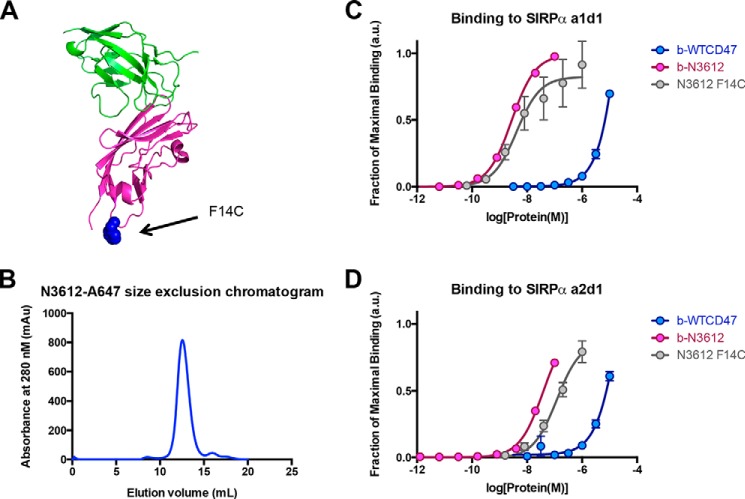
FIGURE 7.
Reformatting high affinity CD47 variant N3612 into a diagnostic agent. A, cartoon representation of x-ray crystallographic structure of the SIRPα (green)-CD47 (magenta) complex. The cysteine mutation (F14C) installed to allow site-specific fluorophore conjugation is highlighted in blue and shown in sphere representation. B, representative size exclusion chromatogram trace of N3612-A647. C and D, titration curves of biotinylated WT CD47 (blue), biotinylated N3612 (pink), and N3612 F14C conjugated to Alexa Fluor 647 (gray) binding to yeast expressing SIRPα a1d1 (C) and yeast expressing SIRPα a2d1 (D). Data represent mean fluorescence intensity normalized to maximal binding for each cell type ± S.E. Data are representative of two independent experiments. a.u., arbitrary units; mAu, milli-absorbance units.

