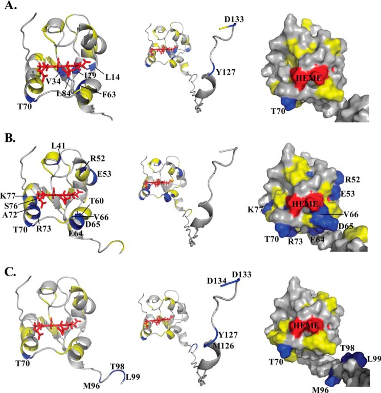
FIGURE 5.
Mapping of differential line broadening of cytb5 residues upon interaction with wt-CYP2B4. Differential line broadening of cytb5 residues is mapped onto the cytb5 structure upon interaction with wt-CYP2B4 at a molar ratio of 1:1 in a lipid-free solution (A), a solution containing DLPC/DHPC isotropic bicelles (B), and a solution containing DPC micelles (C). Residues are categorized and color-coded according to their relative intensities: blue for significantly perturbed residues upon interaction with wt-CYP2B4 with relative intensities more than one S.D. below the average, yellow for moderately perturbed residues with relative intensities in the range from average value to one S.D. below the average, and gray for residues with negligible to no perturbation, of which the relative intensities are above the average value. Heme is colored in red. Significantly perturbed residues are also labeled by amino acid name and the sequence number. Ribbon representations of the soluble domain of cytb5 are presented in the left panel. The active site of cytb5 (front face around the heme edge) is presented in surface representations in the right panel. The full-length structure of cytb5 is shown by ribbon representations in the middle panel to demonstrate the perturbations in the TM domain if present.

