
FIGURE 6.
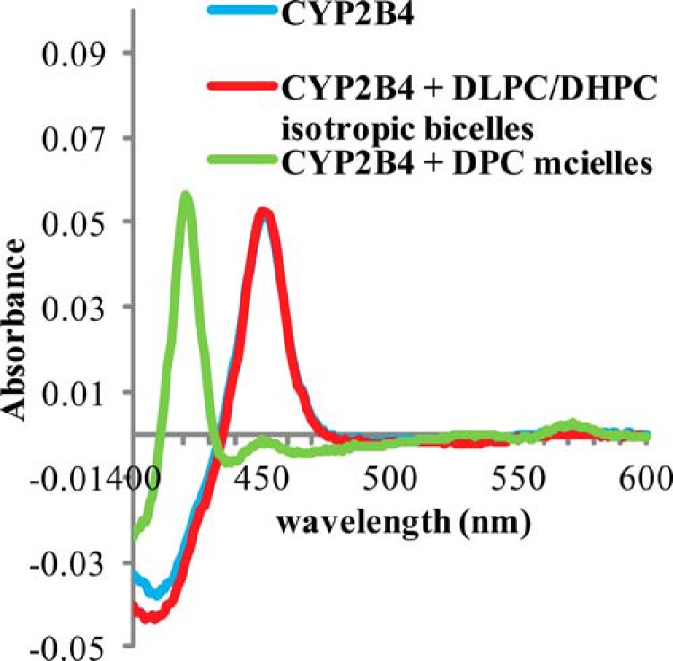
Carbon Monoxide assay of wt-CYP2B4 in different membrane environments. CO assays were performed on 1 μm CYP2B4 at 25 °C in 100 mm potassium phosphate buffer with 5% (w/v) glycerol, pH 7.4, containing no lipid (blue curve) as control, 10% (w/v) DLPC/DHPC isotropic bicelles (red curve), and 2 mm DPC micelles (green curve). An absorption maximum at 450 nm is observed in DLPC/DHPC isotropic bicelle solution, indicative of CYP2B4 in a functionally active P450 form. In DPC micelle solution, CYP2B4 turns into an inactive cytochrome P420 form as shown by a peak at 420 nm in the green curve.