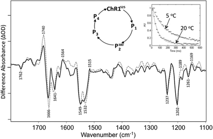
FIGURE 3.
Comparison of time-resolved rapid scan FTIR difference basis spectrum derived from SVD analysis (dashed) recorded at 20 °C and static FTIR difference spectrum (solid) recorded at 270 K. Right inset, SVD-derived decays for time-resolved FTIR difference spectra at 5 and 20 °C. The time-resolved basis spectrum agrees well with the 270 K difference data. y axis markers are ∼0.8 and 0.4 mOD for time-resolved and static difference spectra, respectively. A photocycle scheme is also shown that is typical for ChRs.

