FIGURE 2.

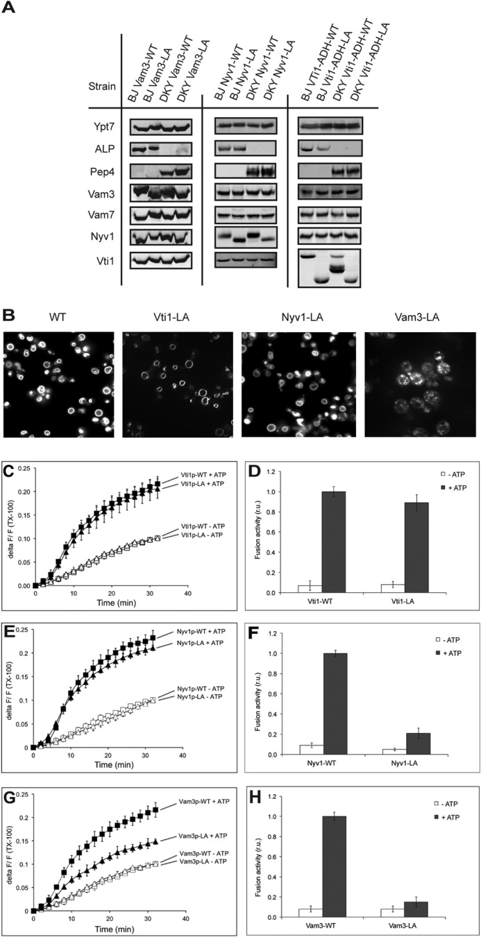
Fusion reactions with vacuoles isolated from Vam3-LA, Nyv1-LA, and Vti1-LA mutants. A, vacuoles were purified from the indicated strains and analyzed by SDS-PAGE and Western blotting for various components of the fusion machinery and for the indicator proteins pro-ALP and proteinase A. B, vacuole morphology of Vam3-LA, Nyv1-LA, and Vti1-LA mutants compared with WT. The cells were grown in YPD and stained with FM4-64, and the structure of vacuoles was analyzed by confocal fluorescence microscopy. C–H, vacuoles were isolated from the indicated strains and used for in vitro fusion reactions in the presence or absence of ATP. Lipid (C, E, and G) and content (D, F, and H) mixing were assayed in parallel. Means ± S.D. are shown for three independent experiments.
