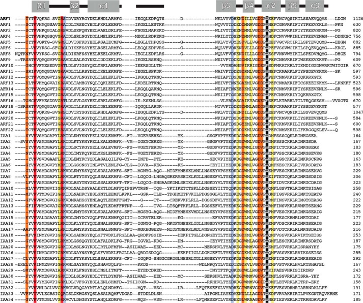
FIGURE 4.
Multiple sequence alignment of PB1 domains from A. thaliana ARF and Aux/IAA. The secondary structure of the Arabidopsis ARF7 PB1 domain (Ref. 13; PDB code 4NJ6) is shown at the top of the alignment. Sequences of the PB1 domains of Arabidopsis ARF and Aux/IAA were aligned using MegAlign (DNASTAR). Colors used to highlight key positions correspond to the effect of alanine mutations on Kd determined by ITC experiments (see Table 3), as follows: light purple, <2-fold; yellow, 2–10-fold; orange, >10-fold. Residues that abolished detectable interactions are red. The structurally important tryptophan is black.

