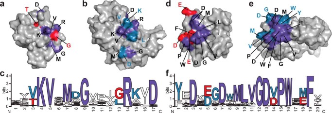
FIGURE 8.

Core and family-specific PB1 domain interface residues. The x-ray crystal structure of ARF7PB1 (Ref. 13; PDB code 4NJ6, chain A) and the NMR solution structure of IAA17 (Ref. 15; PDB 2MUK) are represented as space-filling models with highlighted areas of sequence conservation. Residues conserved in ARF (red), Aux/IAA (blue), and in both ARF and Aux/IAA (purple) are shaded as indicated. a, surface view of the ARF7PB1 basic face. b, surface view of the IAA17PB1 basic face. c, amino acid sequence logo showing conservation of residues on the ARF and Aux/IAA PB1 domain basic faces. d, surface view of the ARF7PB1 acidic face. e, surface-view of the IAA17PB1 acidic face. f, amino acid sequence logo showing conservation of residues on the ARF and Aux/IAA PB1 domain acidic faces. Panels e and f were generated using WebLogo (39).
