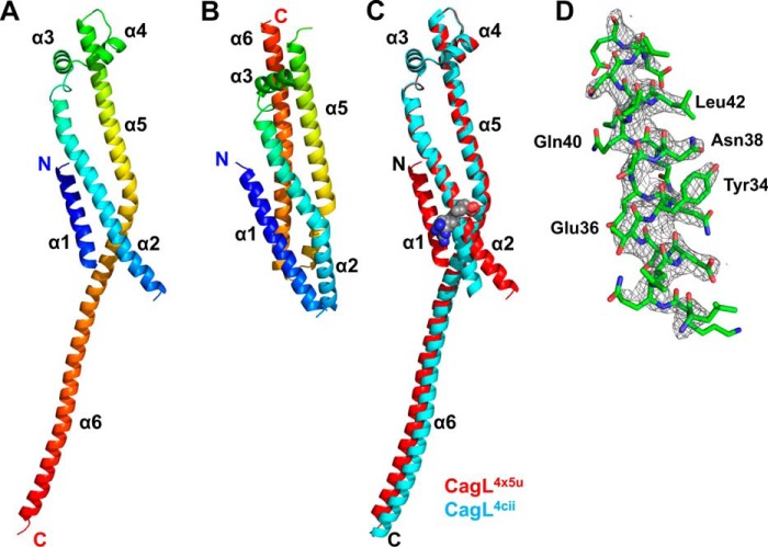
FIGURE 1.
Stability and structure of CagL. A, schematic ribbon of CagL structure solved at pH 4.5 showing the 6 helices from the N terminus (blue) to the C terminus (red). B, structure of the compact form of CagL3zci. C, superposition of CagL4cii (blue) onto the recent crystal structure, CagL4x5u (red). The presence of α1 in the new structure causes straightening of the N-terminal half of α2 below the RGD motif (gray spheres). D, representation of the 2Fo − Fc σA-weighted electron density map contoured at 1.5 σ showing α1 residues of CagL.

