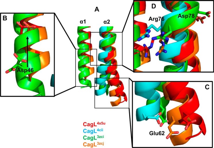
FIGURE 5.
Structural changes of α1 and α2 in CagL in response to pH. A, superposition of all four CagL structures; CagL4x5u, pH 4.5 (red); CagL4cii, pH 4.2 (cyan); CagL3zci, pH 7.4 (green); and CagL3zcj, pH 6.5 (orange). B, lowering of the pH (CagL4x5u and CagL3zcj) causes a registry shift of α1 (CagL3zci) as monitored by Asp46 moving up by one turn of the helix. C, movement of α1 causes the N-terminal half of α2 below the RGD motif to move. With α1 in the up position (CagL4x5u and CagL3zcj), α2 is not kinked and remains straight. When α1 is in the down position or missing (CagL4cii and CagL3zci), α2 becomes kinked. Glu62 shows the degree of movement of α2. D, conformational changes in α1 and α2 result in small differences in the RGD motif in α2. In CagL4x5u, Arg76 forms a salt bridge to α1, whereas in the other structures, Arg76 is either free or the backbone is in a distorted conformation.

