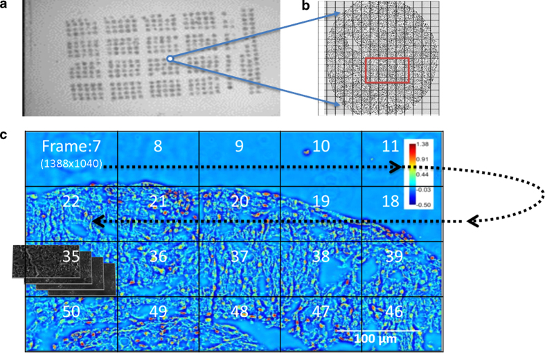
Figure 2. Mosaic SLIM imaging of an unstained tissue microarray.
(A) Unstained tissue microarray slide. (B) The mosaic is set up around the core of interest. (C) The recording at each tile proceeds as shown by the arrow. For each 1388 × 1040 pixel SLIM tile, four intensity images are recorded. The phase images are then stitched together using an ImageJ plugin built in our lab.

