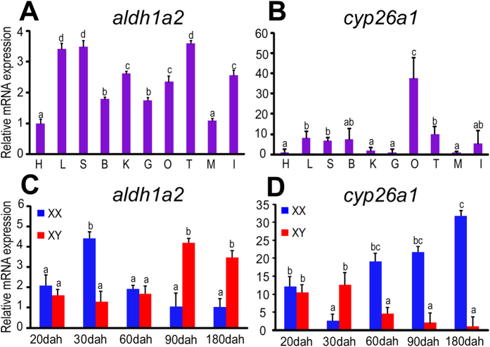
Figure 2. Spatial and temporal expression of aldh1a2 and cyp26a1.
A-B, expression of aldh1a2 (A) and cyp26a1 (B) were detected in adult tilapia (180 dah) tissues. B, brain; G, gill; H, heart; L, liver; I, intestine; S, spleen; M, muscle; O, ovary; T, testis; K, kidney. C-D, expression of aldh1a2 (C) and cyp26a1 (D) were detected during the critical period of germ cell meiosis initiation in tilapia gonads. Data were expressed as mean ± SD (n = 5). Different letters indicate statistical differences at P < 0.05 as determined by one-way ANOVA with a post-hoc test. Dah, days after hatching.

