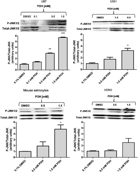
Figure 3.

The effects of POH on the activation of JNK1/2 in U87 and U251 cells, VERO cells and mouse astrocytes. The cells were treated with POH for 30 minutes. The graph shows the densitometric analysis of p-JNK1/2 relative to the total JNK1/2 and is shown in arbitrary units as the ratio of the band densities from western blots of p-JNK1/2 and total JNK1/2 corrected for control. The figures are representative of three independent experiments. The graph represents the means ± SD from at least three different experiments. *p < 0.05, **p < 0.01, ***p < 0.001 vs. control group (0.1% DMSO), analyzed by Student’s t-test.
