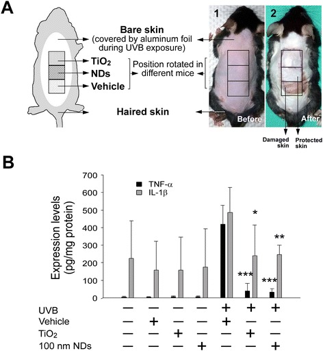
Figure 3.

NDs protect C57BL/6J mouse skin from UVB-induced inflammation. Experiment setting (A) and the typical appearance of hair-removed mouse skin before (A1) and three days after UVB irradiation [UVI 6, 20 min per day, 3 cycles; (135.9 mJ/cm2/day × 3 days)], with or without protection at 2 mg/cm2 nanomaterial density (A2). Bare skin surrounding the experimental area was protected using aluminum foil. Each material was applied to the anterior, middle, and posterior position three times on three mice (A). TNF-α and IL-1β in mouse skin are reported as pg per mg of protein (B). n = 9 (three experiments repeated three times). *P < 0.05, **P < 0.01, ***P < 0.001; significant amelioration versus respective UVB vehicle groups. Data are mean ± SD.
