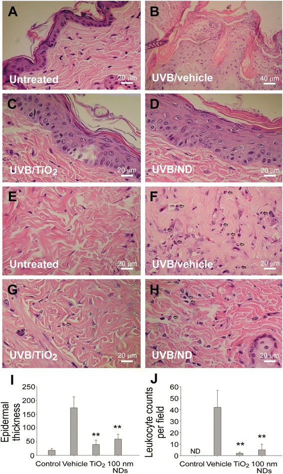
Figure 4.

NDs ameliorate UVB-induced skin hyperplasia and leukocyte infiltration. Histological examinations of the epidermis (A–D) and dermis (E–H) revealed the alterations before (A, E) and 3 days after UVB irradiation [UVI 6, 20 min per day, three cycles; (135.9 mJ/cm2/day × 3 days)], with and without protection using 2 mg/cm2 TiO2 and 100-nm ND nanomaterials (B–D, F–H; H&E stain, × 400, scale bars in A and C–H = 20 μm and in B = 40 μm). The epidermal thickness was quantified as indicated (I). The infiltrated leukocytes (indicated by arrows) were found in the dermis, especially in the vehicle group (F–H); quantified results are presented in (J). n = 9, **P < 0.01, significant amelioration versus vehicle groups. Data are mean ± SD.
