
Figure 5.
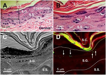
Detecting 100-nm NDs on UVB-irradiated mouse skin. An H&E-stained mouse skin section was used as a sample to illustrate the relative positions and morphology of the epidermis, dermis (A), stratum corneum, and stratum granulosum (S.G.) (B). The area indicated by the black box in (A) is magnified in (B). Differential interference contrast (C) and confocal microscopy (D) images of a 100-nm-ND-shielded UVB-irradiated mouse skin. Dosages of UVB irradiation and 100-nm NDs are the same as those in Figure 4. White arrows indicate the fluorescent signals (501–511 nm; green labels) emitted by the 100-nm NDs, which did not penetrate beyond the S.G. (D). Stratum corneum (S.C.), S.G., stratum spinosum (S.S.), and stratum basale (S.B.) are indicated. Blue arrows indicate keratin autofluorescence.