
Figure 3.
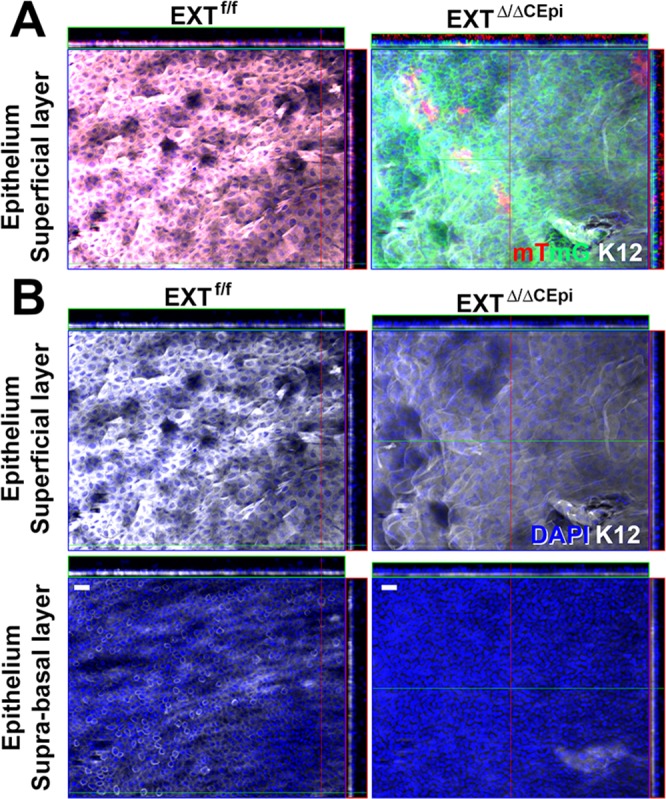
Krt12 staining of corneal whole mounts. Corneas isolated from EXTf/f and EXTΔ/ΔCEpi mice at P55 were analyzed by whole-mount immunostaining. (A) Krt12 (white) staining was colocalized with mTmG representing the cells from which Ext1 was ablated (green), revealing a loss of Krt12 staining in Ext1-null cells. (B) Krt12 (white) staining in the suprabasal and superficial layers of the cornea. EXTΔ/ΔCEpi mice present a loss of Krt12 staining in the suprabasal and wing cell layers and a decrease in the superficial layer of the cornea. Scale bars: 20 μm.