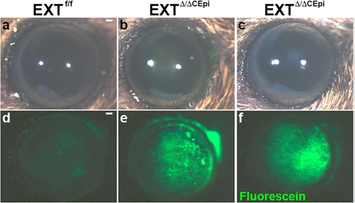
Figure 4.
Loss of barrier function in EXTΔ/ΔCEpi mice. Images were captured of EXTf/f (a) and EXTΔ/ΔCEpi mice (b, c) induced from P21 to P55 and P21 to P120, respectively) prior to fluorescein administration using a stereomicroscope coupled with white light. Fluorescein sodium 0.25% was applied as an eye drop to the eyes of P55 EXTf/f (d) mice, EXT1Δ/ΔCEpi (e) mice induced from P21 to P55, and EXTΔ/ΔCEpi (f) mice induced from P21 to P120. Excess fluorescein was washed off with PBS and images were acquired using the 488 filter set. Scale bar: 250 μm.

