
Figure 5.
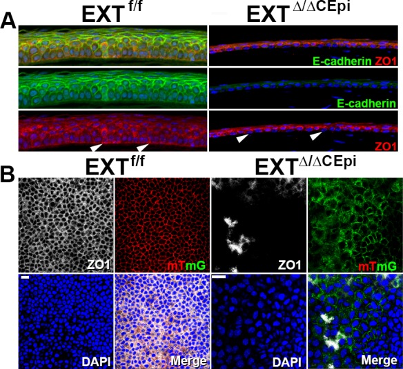
Loss of cell–cell and cell–basement membrane tight junctions in EXTΔ/ΔCEpi mice. (A) ZO-1 (red) and E-cadherin (green) staining was analyzed in sections of EXTf/f and EXTΔ/ΔCEpi mice induced from P21 to P55. ZO-1 staining evidences the loss of adhesion complexes between the basal epithelial cells and basement membrane (arrowheads). (B) Whole-mount analysis was performed to analyze ZO-1 (white) distribution throughout the cornea of EXTf/f and EXTΔ/ΔCEpi mice induced from P21 to P55. Corneas used for whole-mount analysis contained the mTmG gene whereby cells lacking Ext1 present membrane-bound GFP (green), whereas the corneal epithelium of littermate control mice presents membrane-bound tomato red (red). Scale bar: 20 μm.