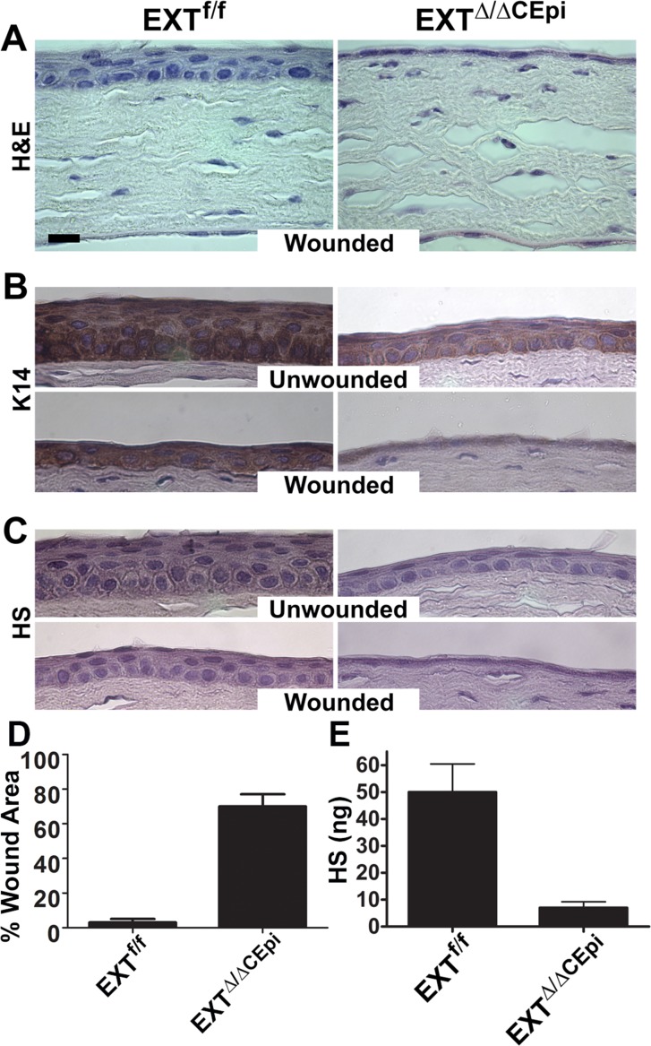
Figure 6.
Wound healing of EXTf/f and EXTΔ/ΔCEpi mice. Debridement wounds were performed on P55 EXTf/f and EXTΔ/ΔCEpi mice induced from P21. The corneas were analyzed after 24 hours. EXTΔ/ΔCEpi mice presented compromised wound healing. Corneas were processed for paraffin sectioning and analyzed by (A) hematoxylin and eosin staining, (B) Krt14 staining, and (C) heparan sulfate staining of the wounded and unwounded contralateral cornea. (D) Percentage of wound area remaining 24 hours after wounding. The corneas of EXTf/f mice healed within 24 hours; however, EXTΔ/ΔCEpi mice had approximately 70% of the wound area remaining 24 hours after wounding. (E) GAGs were extracted from the corneas of EXTf/f and EXTΔ/ΔCEpi mice induced from P21 to P55 and chondroitin sulfate (CS) and dermatan sulfate (DS) was digested by chondroitinase ABC digestion. HS levels were assayed using the carbazole colorimetric assay, revealing a loss of HS expression in the corneas of EXTΔ/ΔCEpi mice when compared to littermate control mice (EXTf/f). Scale bar: 20 μm.

